
What is Yellow Body Cyst?
Cyst of the yellow body occurs due to the accumulation of fluid in the place of the bursting follicle, sometimes may contain blood. Such cysts occur only during a biphasic menstrual cycle. It is believed that these cysts are formed as a result of impaired lymph and blood circulation in the corpus luteum. Occur in age from 16 d to 45 years.
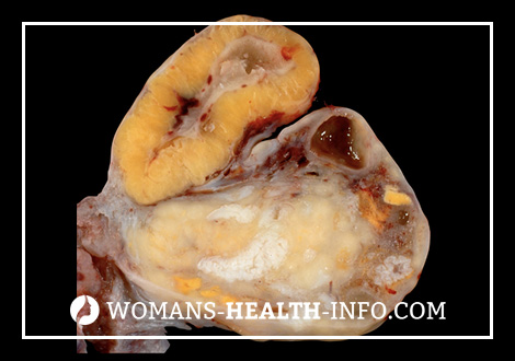
Pathogenesis during Yellow Body Cyst
Microscopically, lutein and theca-lutein cells are detected in the wall of the cyst of the corpus luteum. Luteal cells undergo all stages of the development of the menstrual yellow body – proliferation, vascularization, flowering and reverse development.
Symptoms Yellow Body Cyst
Clinically, a cyst usually does not manifest itself. Rarely disrupted menstrual cycle. Specific clinical signs are absent. In some cases, at the time of occurrence of a cyst, pain in the lower abdomen may occur.
The most frequent complication is hemorrhage into the cyst cavity. Cysts of the corpus luteum are often complicated by rupture of the ovary, often in the stage of development of the corpus luteum. Bleeding can be intense and is accompanied by a clinical picture of the “acute abdomen.”
In most cases, cysts of the corpus luteum undergo the opposite development. The luteal cell layer is gradually replaced by connective tissue and the formation can turn into a cyst, the inner surface of which is devoid of the epithelial lining.
Diagnosis Yellow body Cysts
The diagnosis of cyst of the corpus luteum is established on the basis of anamnestic data, the results of clinical examination, ultrasound and DDC. laparoscopy.
In a two-handed vaginal-abdominal examination, the corpus luteum cyst is located mainly laterally or posteriorly from the uterus, round in shape, with a smooth surface, elastic consistency, with a diameter of 3 to 10 cm, as evidenced by ultrasound data, mobile, sensitive to palpation.
The echographic picture of cysts of the corpus luteum is very diverse. The cyst structure can be completely homogeneous and anechoic or have a small or medium-mesh structure, and these structures can perform the entire cyst or a small part of it. In the cavity of the cyst are determined by multiple partitions of irregular shape, shifted by the formation of percussion, ultrasonic sensor. In some cases, dense inclusions of increased echogenicity — blood clots — are visualized in the cyst cavity. On scans, wall-mounted inclusions with a diameter of up to 1 cm of irregular shape are visualized; in individual observations, a dense formation is suspended in the cyst cavity. Sometimes the entire cavity of a cyst is filled with echogenic contents (blood), and therefore the sonographic image resembles a tumor. Despite significant differences in the internal structure of cysts of the yellow body, their sound conductivity is always high.
Cysts of the yellow body disappear within 1 – 3 menstrual cycles, which is confirmed by ultrasound data.
Color Doppler allows to exclude the point of vascularization in the internal structures of cysts of the corpus luteum and thus conduct a differential diagnosis with ovarian tumors.
The cyst of the corpus luteum has an intensive blood flow around the periphery (the so-called coronary) with low vascular resistance (IL below 0.4), which often resembles malignant neovascularization.
Treatment for Yellow Body Cyst
To eliminate errors, it is necessary to have a dynamic ultrasound scan with a DDC in the first phase of the next menstrual cycle. In cysts of the corpus luteum, observation is observed during 1-3 menstrual cycles, since its reverse development is not excluded. Otherwise, surgical treatment is indicated – removal (enucleation) of a cyst within the healthy tissue of the ovary with laparoscopic access. Retention cysts are usually small, with a thin transparent wall through which homogeneous contents appear through. With laparoscopy, several small cysts can be seen. With lateral illumination, the retention formations acquire a uniform bluish tint. For small retention cysts, the unchanged surface of the rest of the ovary with follicles or yellow bodies is visualized. The vascular pattern of a capsule can be diverse, but is usually presented as a looped net.
The possibilities of ultrasound with a CDC, CT, MRI for retention formations in the description of their shape, size, structure and location are the same, and when using techniques with contrasting retention formations do not accumulate a contrast agent, which is a differential diagnostic sign of the cyst, indicating retentional mass education.
The prognosis is favorable.