
What is Pregnant Nephropathy?
Nephropathy of pregnant women, or late pregnancy toxicosis, is a disease that occurs in women with healthy kidneys, usually in the third trimester of pregnancy and after it ends. Such nephropathy is called primary. It manifests itself in proteinuria, edema and hypertension, and both mono- and polysymptomatic toxicosis are possible. Among the causes of maternal and perinatal infant mortality, nephropathy of pregnant women is a relatively high proportion. The frequency of nephropathy of pregnant women, according to different authors (I.P. Ivanov, 1971; N. B. Sobenin, 1978), ranges from 2.2-15.0%.
Complex or combined toxicosis is distinguished, which develops in pregnant women with the presence of pre-existing glomerulonephritis, pyelonephritis and other kidney diseases, as well as in hypertension, heart defects, and especially aortic insufficiency, which occurs with high blood pressure. Such toxicosis is also called secondary. For the mother and the fetus, the risk of undesirable consequences in this case increases significantly.
Causes of Pregnant Nephropathy
There are many attempts to explain the development of late toxicosis from different perspectives. Some hypotheses recognize the occurrence of harmful metabolic products in the ischemic uterus and placenta as a decisive factor. It is believed that the ischemic placenta produces vasopressor substances (hysterotonin, etc.) that cause a generalized spasm of arterioles, or toxic metabolic products are formed in it, among which proteolytic enzymes are present. The latter are antigens. The resulting antigen-antibody complexes, settling in the kidneys, damage the renal glomeruli. It is also possible that thromboplastins come from the ischemic placenta into the general circulation, provoking the development of disseminated intravascular coagulation syndrome (DIC). Repeated thromboembolism of the kidneys, lungs, and a large circle of blood circulation support this assumption.
From the hypotheses of the second group, it follows that the development of nephropathy in pregnant women is based on a violation of hormonal homeostasis. Metabolites formed in the ischemic uterus and placenta stimulate the secretion of adrenal hormones, in particular the mineralocorticoid aldosterone and the pressor substances of the brain substance – catecholamines. This leads to an imbalance between aldosterone and progesterone in favor of the former. In addition, the production of renin increases not only in the kidneys, its extrarenal synthesis occurs in the placenta and uterus. The placenta synthesizes important blood flow regulators – prostoglandins, while in pregnant women with nephropathy they find an active mediator of vasoconstriction – serotonin.
Pathogenesis during Nephropathy of Pregnant Women
An important role in the pathogenesis of nephropathy in pregnant women is given to the immunological conflict between the mother and the fetus with the formation of immune complexes containing IgG, IgM, as well as the C3 complement fraction. This immunological conflict is one of the triggers of late toxicosis. In the mother’s body, reactions develop with the release of biologically active substances – acetylcholine, serotonin, heparin, histamine, etc. These immunological, neurohumoral disorders first of all lead to a breakdown of the mechanisms responsible for the functional state of the circulatory system.
The following changes occur in the patient’s body: generalized spasm of blood vessels at the level of arterioles and arterial knees of the capillaries, expansion of veins, increased permeability of the vascular wall, redistribution of fluid, activation of plasma and cellular units of hemostasis, impaired state of blood aggregation. As a result, circulatory and histotoxic hypoxia develop, leading to a violation of the functions of vital organs.
In the occurrence of late toxicosis of pregnant women, an important role belongs to the dysfunction of the central nervous system. This is evidenced by deviations in the central nervous system, established on the EEG long before the onset of clinical symptoms of the disease.
The many hypotheses about the causes of toxicosis of pregnant women and the mechanisms of its development indicate that this issue continues to be debatable.
Morphologically detect a generalized spasm of arterioles, obliteration of their fibrin microtrombi and intravascular aggregation of blood cells. Significant changes are found in the kidneys. The glomeruli are enlarged, ischemic, the walls of the glomerular loops are thickened, swollen, the intracapsular space is narrowed, and fibrin deposits are found in it. Bringing glomerular arterioles are edematous and severely spasmodic. Identified tubular changes, mainly in the proximal sections, are of varying degrees of severity: from dystrophic changes in the epithelium to the development of tubular necrosis.
Using electron microscopy, signs of nephropathy are established, such as narrowing of the glomerular capillary lumen with endothelial cell hyperplasia; thickening in some places of the basement membrane. Consequently, morphological changes in the kidneys during nephropathy of pregnant women are similar to membranous or membrane-proliferative glomerulonephritis and differ from it only in a large lesion of the arterioles, more pronounced dystrophic changes in the epithelium of the tubules and juxtaglomerular cells. The indicated changes in the kidneys with nephropathy are reversible and after delivery (according to the puncture biopsy) in most cases quickly disappear.
With nephropathy of pregnant women, changes in the liver, myocardium, and blood vessels of the brain are possible.
Symptoms of Pregnant Nephropathy
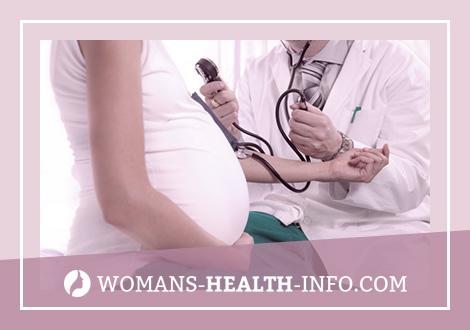
The main clinical manifestations of nephropathy in pregnant women are edema, hypertension, proteinuria. The classic “triad” of nephropathy symptoms is observed in about 50-60% of patients. In other cases, late toxicosis is characterized by two or even one symptom. However, these toxicosis variants are no less dangerous than classical nephropathy. The most frequent and important manifestation of nephropathy in pregnant women is hypertensive syndrome. If a woman in the second half of pregnancy, blood pressure exceeds 130/85 mm RT. Art. or increases by 20-30 mm RT. Art. compared to the original, nephropathy should be suspected. At the same time, it is taken into account that in healthy women, during normal pregnancy, blood pressure remains almost unchanged. Hypertension can be significant, but rarely goes into malignant form. She is a kind of harbinger of eclampsia. High hypertension can cause overload of the left ventricle with symptoms of cardiac asthma and pulmonary edema.
The development of late toxicosis should be considered very unfavorable against the background of hypertension before pregnancy, since in such cases its course is more severe. In addition, there is a risk of underestimating the initial pressure. A poor prognostic sign is a high level of diastolic pressure, even with relatively low systolic pressure.
Complaints in pregnant women with late toxicosis may be absent, but in most cases they are concerned about headaches, irritability, increased fatigue, and visual impairment.
Changes in the vessels of the fundus are not always observed. More often they are similar to those that occur with hypertension – spasm of arterioles (hypertonic angiopathy), swelling of the optic nerve, hemorrhage and foci of degeneration. In severe cases (with malignant hypertensive syndrome), pronounced changes in the fundus are an indication for urgent delivery. Nephropathy of pregnant women is characterized by the disappearance of changes in the fundus with normalization of blood pressure. On the contrary, severe and persistent changes in the fundus often indicate chronic nephritis, hypertension.
The second most common occurrence is edematous syndrome. Initially, edema is insignificant and objectively difficult to determine. Therefore, a weekly weighing of a pregnant woman is mandatory. An increase in body weight of more than 600 g per week indicates a pathological fluid retention in the body. First, swelling appears on the legs, then spreads to the hips, lower back, abdomen, mammary glands, less often on the face. Cavitary edema is rare. Diuresis is usually reduced, and with significant edema, especially rapidly developing, pronounced oliguria can be observed.
Proteinuria, reaching 1-6 g / l, and sometimes 40 g / l or more, in combination with microhematuria and cylindruria is the third important clinical and laboratory sign of pregnant nephropathy. More significant hematuria may indicate a combination of nephropathy with glomerulonephritis.
Renal function with pure toxicosis is not significantly impaired: the concentration ability of the kidneys, blood levels of urea and creatinine are within normal limits. Only in severe toxicosis with severe oliguria or anuria, a transient decrease in renal blood flow, glomerular filtration, and moderate hyperazotemia can be noted.
In cases involving liver damage, pain in the right hypochondrium and enlargement of the liver are observed, sometimes jaundice appears. At the same time, there is a violation of the prothrombin-forming, detoxification, protein-forming function of the liver. In the latter case, albumin deficiency and dysproteinemia are clearly expressed; increased content of lipoproteins, cholesterol, sugar; ESR accelerated.
Vascular disorders worsen the activity of the heart muscle, causing the development of the so-called “ischemic myocardiopathy” observed in severe forms of late toxicosis. Along with this, changes in the blood coagulation system, water-salt metabolism with a delay in sodium and water, inhibition of the function of the thyroid and pancreas are noted. In practice, mild forms with unexpressed clinical manifestations currently prevail among late toxicosis.
Diagnosis of Pregnant Nephropathy
In differential diagnosis, it is necessary to keep in mind various kidney diseases (glomerulonephritis, pyelonephritis). In such cases, a more severe course is noted, toxicosis occurs earlier in pregnancy, is difficult to treat, and leads to a significant increase in perinatal mortality. In this case, the time of occurrence of nephropathy, the absence or presence of a history of kidney disease should be taken into account.
The latent course of glomerulo- or pyelonephritis often creates a false idea of the timing; its occurrence. Therefore, for the purpose of timely diagnosis and treatment, a systematic urinalysis and measurement of blood pressure in all pregnant women at the earliest possible time are necessary. In addition, the study helps the functional ability of the kidneys, whose changes in primary nephropathy are not pronounced. The most effective means of early diagnosis is the medical examination of pregnant women. It involves the systematic and careful monitoring of a pregnant woman in consultation and during patronage visits (monitoring the pregnant woman’s weight, measuring blood pressure, urinalysis, identifying previous kidney diseases and all diseases predisposing to nephropathy).
Complications With the correct and timely treatment of nephropathy of pregnant women, the prognosis is favorable. Recovery can go in two ways. The first one lasts a few days after giving birth, the second one is longer, up to 1.5 months. During this time, edema disappears, then hypertension, proteinuria decreases and soon disappears, partial kidney function is restored.
In the renal tissue (with a biopsy) within the specified time after childbirth, as a rule, pathological changes that have taken place are not detected.
In severe late toxicosis, in cases of preeclampsia, the clinical picture consists of the symptoms of acute malignant hypertension. Pronounced headaches, nausea, vomiting, loss of appetite, often loss of vision prevail. Mental disorders (stiffness, lethargy), acute heart failure can occur. Along with this, edema is observed, often massive, high proteinuria. Preeclampsia is a transitional stage to a more serious complication of pregnant nephropathy – eclampsia, which occurs in about 1.5% of cases of pregnant nephropathy and is characterized by the addition of tonic and clonic seizures to the described clinical picture, loss of consciousness.
The mechanism of eclampsia is similar to the mechanism of hypertonic encephalopathy in acute glomerulonephritis (a sharp increase in intracranial pressure, cerebral edema). Each attack of eclampsia begins with a small twitch of the muscles of the face, eyelids, then convulsions of the entire skeletal muscles (tonic) develop, finally there is a violent convulsive twitch of the muscles of the face, trunk, upper and lower extremities (clonic convulsions). The development of eclampsia is often accompanied by fever, respiratory arrest, cyanosis. During the resolution of the attack, a coma develops with a gradual return of consciousness. The duration of the seizures is 30-40 s. During the day, they can often be repeated so that the patient practically does not have time to regain consciousness. Occasionally, a patient falls into a prolonged coma without previous seizures. This is the most severe and dangerous form of eclampsia.
Statistics show that eclampsia attacks can occur before childbirth in 25%, during childbirth in 50 and after in 25% of cases. The outcome of an attack is determined by the level of blood pressure and the degree of cerebrovascular accident. Mortality in eclampsia is 1-9% and occurs from cerebral hemorrhage or from acute heart failure. A spasm of the renal arterioles during an attack can cause tubular necrosis and acute renal failure.
Diagnosis of eclampsia is usually not difficult, but in some cases it is necessary to be able to distinguish it from a diabetic and uremic coma, Morgagni-Adams-Stokes syndrome. In 3.4% of cases (K. N. Zhmakin, 1979), eclampsia recurs during subsequent pregnancies. Nephropathy complicated by eclampsia causes persistent consequences: in 1/3 patients there is a violation of cerebral circulation, decreased vision and other changes, in 20% later there are deviations from the side of kidney function, up to the development of chronic renal failure. In 17.9% of patients, pregnant nephropathy is transformed into hypertension (V.V. Razumov, 1983).
Pregnant Nephropathy Treatment
With nephropathy of pregnant women, first of all, a gentle regimen is needed. In severe cases of nephropathy, they are shown bed rest, diet and medication.
As a rule, treatment of nephropathy of pregnant women is carried out inpatiently in specialized departments (pathology of pregnant women). A mandatory component of treatment is diet (table No. 7). The main requirements for it are as follows: to limit the daily intake of salt (up to 1.5-3 g), especially with high hypertension and preeclampsia, and fluid (up to 1 liter). Consumption of the latter is distributed in equal portions. The amount of protein in the daily diet remains normal (1-1.2 g per 1 kg of body weight, including half of it must be of animal origin). The amount of fat decreases slightly and amounts to 0.7-1 g per 1 kg of body weight. A sufficient amount of foods rich in carbohydrates and potassium must be introduced into the daily diet. Alcoholic beverages are contraindicated. Fasting days are recommended once every 7 days (cottage cheese, dried fruit, etc.).
Of medications, sedatives are immediately necessary. This allows you to normalize the activity of the central nervous system. In the future, medications are prescribed based on the symptoms of toxicosis. So, to eliminate hypertension, individually selected antihypertensive drugs of all groups can be recommended. It is advisable to use drugs with a different mechanism of action: antispasmodics, adrenergic blockers, peripheral vasodilators (aminophylline, papaverine, dibazole, pyroxan, obzidan, adelfan, methyldopa, apressin) (A. Yu. Nikolaev, V.A. Rogov, 1989).
Only the use of guanidine derivatives (isobarin, ismeline) is contraindicated, since these drugs can cause orthostatic collapse in a pregnant woman, severe complications and even fetal death.
To eliminate edema and increase diuresis, diuretics are used in different combinations simultaneously or sequentially. The purpose of diuretics is combined with a sufficient intake of potassium. Spironolactones may be recommended. With all forms of nephropathy, intravenous or intramuscular infusions of aminophylline, magnesium sulfate are prescribed in parallel.
With the development of preeclampsia and eclampsia, the fight against cerebral edema is of paramount importance. The classic remedy is 20 ml of a 10% solution of magnesium sulfate intravenously, then 10 ml of a 25% solution intramuscularly. Parenteral diuretics (lasix) are prescribed. You can enter reopoliglyukin, mannitol, 40% glucose solution, glucose-novocaine mixture. As an osmotic diuretic, glycerin is used at 0.5 g / kg body weight 2 times a day along with fruit juice. Antipsychotics (droperidol), seduxen, barbiturates, chlorpromazine, chloral hydrate in enemas are indicated. If eclampsia attacks do not stop, promedol or pipolfen are administered intravenously.
In addition to the listed traditional methods of treating severe toxicosis, patho-
genetic agents. Positive results were obtained using anticoagulants (direct and indirect), antiplatelet agents. Pregnant women with eclampsia are given concentrated plasma, a 20% albumin solution. During treatment, constant monitoring of acid-base balance, blood coagulation and the functional state of the kidneys is necessary. To normalize immunological disorders in severe toxicosis, thiol preparations are used (a course of intramuscular injections of unitiol). If there is no effect with conservative therapy, an urgent delivery is performed.
Prevention of Pregnant Nephropathy
Since many women who have experienced severe forms of nephropathy in pregnant women experience changes in their urine and hypertension in the postpartum period, they must be taken to the dispensary. The minimum period of medical examination is a year during which patients undergo treatment and control at least once every three months under the supervision of a local physician and nephrologist. After that, depending on the results obtained, the question of the termination or extension of dispensary observation is decided. In the latter case, the approach to clinical examination should be twofold. If the patient has signs of renal pathology, she should undergo the same treatment and control as patients with glomerulonephritis. If hypertensive syndrome persists, appropriate treatment by a local GP or cardiologist.